 |
 |
- Search
| Neonatal Med > Volume 30(4); 2023 > Article |
|
Abstract
The word “microbiome” is a combination of “microbiota” and “genome,” which represents the genomic concept of microbiota. The bacterial culture method is the mainstay of identifying microbes, while polymerase chain reaction adds diagnostic value. However, in the era of next-generation sequencing, achieving high-throughput microbiota footprints is extremely sensitive. This sensitivity often leads to confusion, as it can detect specific microbes genomes, even in sterile samples, such as blood, placenta, breast milk, skin, vagina, and stool. The neonatal microbiome remarkably influences both fetal and neonatal life related to health status and disease outcome. However, its origins pose a question: does it stem from a direct gateway or through a breakdown of barriers? This review provides a brief overview of evidence and speculative insights.
인류는 인체 내에 공생하는 미생물과 함께 진화해 왔고, 인체 내 미생물이 인류의 건강에 미치는 영향은 광범위한 것으로 생각되어 진다[1,2]. 그 중에서도 최근의 연구들이 보고하는 바에 의하면 몇 가지 장내 마이크로바이옴의 집락화 패턴과 특정 질환과 연관 짓고 있다. 예를 들면, 염증성 장질환과 관련하여 Bacteroidetes, Bifidobacterium, Clostridium, Lactobacillus가 감소하고 Fusobacterium, Escherichia coli 등은 반대로 증가하는 특징 등이다[3-7]. 이와 같이 제2의 인간 게놈이라고 불리기도 하는 마이크로바이옴이 사람의 건강과 질병에 관여하기도 하면서 세대를 바꾸어 진화와 계승을 반복하는 과정에는 음식물의 섭취, 생활 습관, 환경적 노출의 변화가 중요한 역할을 한다[8]. 그렇다면 신생아가 태어나고 장내 세균이 집락화 하는 과정에서도 환경적 노출이 가장 큰 역할을 할까? 이 질문에 가장 확실한 대답은 아마도 질식 분만 신생아와 제왕절개 출생 신생아의 차이[9-13], 그리고 모유 섭취 여부에 따른 신생아 장내 세균 집락화의 차이가 될 것이다[14-17]. 전통적으로 출생 전의 태아의 모든 조직은 무균적 환경이라고 여겨졌기 때문에, 양수내 감염을 포함한 선천성 감염을 제외한다면, 신생아에서 마이크로바이옴이 집락화를 시작하는 시점은 분명히 출생하는 것이 될 것이고, 그다음으로 모유 섭취가 영아 초기의 장내 세균 확립에 지대한 역할을 하는 것으로 되어 있다.
그럼에도 불구하고 출생 직후를 시작으로 하는 것이 아니라, 출생 이전 즉, 태반을 통로로 해서 산모의 장내 마이크로바이옴이 태아로 전달되는 경로에 대한 연구들이 최근 꾸준히 대두되고 있어 이에 대해 살펴보고자 한다.
먼저, 신생아 및 주산기 마이크로바이옴의 중요성에 대해 살펴보기로 하겠다. 인체의 마이크로바이옴에 대한 연구를 검색하면 2022년 기준으로 2000년에 게재된 연구의 278배에 달하는 만큼 엄청난 속도로 많은 연구들이 쏟아져 나오고 있다(Figure 1). 과거 산발적이었던 연구에 대조적으로 2007년에 National Institute of Health (NIH) 주관으로 시작된 Human Microbiome Project 1 (HMP-1)이 이정표가 되어 본격적으로 많은 연구들이 나오기 시작했는데, 여기에는 주로 인체의 주요 부분들에 어떤 마이크로바이옴들이 분포하고 있는지를 규명하는 것이 주된 목적이었다. 그리고 2014년 HMP-2에서부터는 인체의 미생물들이 사람의 건강과 질병 상태 사이에 어떠한 역할을 하는 지가 규명해야 할 과제가 되었으며 세 가지 주제가 집중적으로 대두되었고 아래와 같다.
1. 조산(preterm birth)
2. 염증성 장질환(inflammatory bowel disease)
3. 당뇨병 전단계
이렇듯 집중 과제 중 하나를 차지할 만큼 마이크로바이옴 연구에 있어 조산은 매우 중요한 영역이며, 주산기 마이크로바이옴 연구에 많은 연구비가 집중되고 있는 것이 사실이다.
전통적으로 자궁 안 환경은 무균적일 것이라는 패러다임이 있어왔다. 즉, 배아로부터 발달한 태아는 파열되지 않은 양막 안에서 발달을 지속하는 한, 외부로부터의 세균이 침입할 근거가 없다는 것이다. 따라서, 태아가 출생하는 과정에서 질식 분만을 하는 경우, 질 내 산도의 분비물을 섭취하기 시작하면서, 혹은 제왕절개술의 과정에서는 양막이 파수 되고 외부 환경의 물질에 노출되기 시작하면서 비로소 아기의 구강에서부터 대장으로 이어지는 장관에서 집락 형성이 이루어지는 것으로 생각되어져 왔다. 그러나, 최근에 이러한 ‘무균적 자궁 가설(Sterile womb hypothesis)’의 개념에 의구심을 제기하는 연구자가 많다. 즉, 세균의 집락화는 이미 정상 자궁 내에서 태아 시기에서부터 시작한다는 가설을 주장하는 연구자들이 많아 지고 있는 것이다[18].
만약 태아의 장관 내에서는 절대적으로 무균이었다면, 출생 직후에 아기의 첫 대변에서는 검출되는 마이크로바이옴이 거의 없어야 할 것이며, 시간이 흐르면서 차츰 개체수나 다양성 면에서 증가하는 경향을 보여야 하는 것이 맞을 것이다. 그러나 2016년에 게재된 체계적 고찰 문헌을 보더라도[19] 이미 신생아의 첫 태변에서부터 다양한 마이크로바이옴이 검출되는 것을 알 수 있듯이 최근의 연구들에서는 오히려 첫 태변에서부터 다양성이 큰 마이크로바이옴 패턴이 분석되고 있을 뿐만 아니라 첫 태변에서의 다양성이 훨씬 높고, 이후 점진적으로 다양성이 감소한다는 연구들이 있기 때문에, 태아의 무균설은 점차 설득력을 상실하고 있다[20-22].
그렇다면 태반에는 과연 마이크로바이옴이 존재할까? 태반 검체 분석을 통해 얻은 마이크로바이옴 정보는 산발적으로 보고되었다. Zakis 등[23]은 최근에 57개의 연구에서 이에 관한 체계적 문헌 고찰을 게재하였는데, 가장 많은 수에서 보고된 태반 조직의 세균 문(bacterial phylum)으로는 Firmicutes로 18개의 연구에서 공통으로 분석되었고, 세균 종(bacterial genus)으로는 Lactobacillus가 11개의 연구로 가장 많이 분석되고 있다.
태반 분석에서 마이크로바이옴이 검출된다고 해서 태반에 세균이 교통하고 있다는 가설의 직접적인 증거가 될까? 태반은 대표적인 낮은 농도의 생질량 조직이다(low bio-mass tissue). 즉, 정상적으로 무균으로 여겨지는 조직으로서 마이크로바이옴의 존재를 분석하기에 매우 어려운 조건을 가지고 있다. 현재의 차세대 서열 분석(next generation sequencing, NGS) 기반의 16s rRNA 분석을 기본으로 한 유전자 분석은 미생물의 유전자 서열 중에서 가변성 구간인 V3–V4의 서열을 분석함으로써, 라이브러리에 등록되어 있는 세균 정보와 합치하는 것을 마이크로바이옴으로 분석한다. 그러나, 이러한 서열 분석을 통하여 얻은 결과를 통해 도출되는 결과 즉, 데이터 은행의 라이브러리 데이터와 합치 배정된 마이크로바이옴의 분석 결과가 반드시 세균 미생물의 온전한 존재와 동일한 의미인지는 아직도 확실하지 않다. 더욱이, NGS 기법의 유전자 분석 도구는 매우 민감하기 때문에, 분석하고자 하는 검체와 함께 분석 기계에도 달한 약간의 오염균, 혹은 오염된 유전자가 존재하더라도 이것이 마치 그 검체에서 유래된 것으로 오해하기 쉬운 결과를 받아 보게 된다. 한 실험적 연구에서는 실제 검체를 포함하지 않은 분석액만으로 서열 분석을 시행했을 때도 일정 부분의 서열 증폭을 보고한 바가 있으며[24], 검체 분석을 위한 검사 도구 자체에서 유래한 오염 유전자를 ‘Kitome’, 분석 용액이 인근 검체로 튀어 에러를 일으켰다고 하여 ‘Splashome’이라고 명명한 연구자들도 있을 정도이다[25,26]. 실제 Olomu 등[26]의 연구에 의하면, 검체를 첨가하지 않은 접시에서조차 Proteobacteria 및 Firmicutes 문(phylum)이 우세종으로 분석되어 나옴으로써 생질량이 적은 검체에서의 메타지노믹스 분석(metagenomics sequencing) 이 타당한 것이냐에 의구심을 제기하였다. 왜냐하면 검체에 미생물 유전자가 오염되어 있다면, 강력한 서열 분석 능력에 의해서 미생물 메타지노믹스 구성 분율에 심각한 왜곡을 야기할 수 있기 때문이다. 그러나, 검출률이 낮다고 해서 마이크로바이옴 연구의 결과들을 모두 인정하지 않을 것인가에 대해서도 풀어야 할 과제인 것은 분명하다.
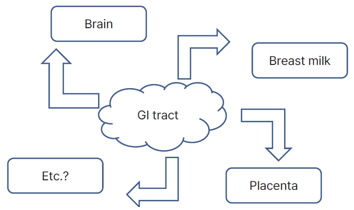
최근의 한 연구에 의하면, 17쌍의 모체-신생아의 219개의 검체를 분석하였는데, 태반, 피부, 구강, 대변, 질, 태변 검체를 포함하여, 태반에서 검출되는 마이크로바이옴과 태변의 것과 비교해 보았는데, 상당 부분 상이한 패턴을 보여 주었다[27]. 물론, 태반의 수가 20개로 통계적 의미를 갖기는 어려웠지만, 향후 이와 같은 대규모의 연구를 통해 의미 있는 전달의 증거를 찾을 필요가 있어 보인다. Aagaard 등[28]이 수행한 연구에 의하면, 16s rRNA 메타게놈 분석을 통해 320명의 산모의 검체를 분석한 결과, 태반 조직에서 검출된 마이크로바이옴으로는 Firmicutes, Tenericutes, Proteobacteria, Bacteroidetes, Fusobacteria phyla 등이 분석되었고, 피부, 구강, 질, 대변에서 분석된 마이크로바이옴과 중에서는 질 유래 마이크로바이옴과는 큰 차이를 보이며, 오히려 구강 마이크로바이옴에서 유사성이 가장 컸다. 물론 이 연구의 제한점으로 구강 내 질환을 구분하지 않은 것이 있지만, 적어도 구강을 위시한 장관 마이크로바이옴이 림프관을 교두보로 하여 혈류를 거쳐 태반 및 제대혈을 거쳐 태아에게 전달될 수 있다는 가설의 근거로 종종 이 연구가 거론된다. 이렇듯 혈행으로 이동하여 해당 장기에 영향을 미친다는 대표적인 가설에는 태반 외에도 장-유선 축(gut-mammary axis), 장-뇌 축(gut-brain axis) 가설이 있다(Figure 2).
모유를 분석한 많은 연구과 체계적 고찰 연구에서 마이크로바이옴의 존재는 확인되고 있다[29,30]. 그러나 그러한 분석에서조차 피부 정상균 등에 의해 오염된 결과일 것이라는 주장도 여럿 존재한다. 예를 들면, 영아가 모유를 먹을 때, 흡철하는 과정에서 아기의 구강에서 유선 쪽 방향으로 역류가 존재한다는 증거에 대해 연구된 바가 있다[31]. 특히 모유 마이크로바이옴이 영아의 장관보다는 구강의 마이크로바이옴과 연관이 있다는 연구에 비추어 보면, 모유 마이크로바이옴은 그 고유의 것이라기보다는 아기 구강 마이크로바이옴에 의해 오염되었을 가능성도 설득력이 없는 것은 아니다[32]. Hassiotou 등[33]은 모유에서 분석된 염증 사이토카인과 수유모 및 아기의 감염과의 관계에 대한 흥미로운 연구를 수행하였는데, 흥미로운 것은 모유의 사이토카인이 아기의 감염 질환 상태에도 반응한다는 것으로 보아 역시 같은 맥락에서 이해할 수 있는 부분이다. 또한, 모유에서 분석되는 마이크로바이옴으로는 대부분의 연구에서 Streptococcus, Staphylococcus, Corynebacteria, Propionibacteria 등이 보고되고 있는데, 이는 피부 상재균이기도 하기 때문에, 이 역시 유선 등에서 유래하는 모유 고유의 마이크로바이옴이라고 하기에는 설득력이 부족한 현실이다.
그렇다 하더라도, 많은 수의 연구들이 모유의 마이크로바이옴에 연관되어 나오고 있다. 특히, 모유의 마이크로바이옴의 유래가 앞서 언급한 것과 달리 혈행(blood stream)을 타고 유선으로 전달되는 경로로서 장-유선 축(gut-mammary axis)의 가설이 설득력을 얻고 있다. 그 증거의 예를 들면, 장관 연관된 절대 혐기성 세균(obligate anaerobes)인 Bifidobacterium, Bacteroides, Parabacteroides, Clostridia (Blautia, Clostridium, Collinesella, Veillonella) 들이 수유모의 대변, 모유, 그리고 신생아의 대변에서 공통으로 도출된 것을 볼 수 있다[34]. 모유를 검체로 해서 마이크로바이옴을 분석하는 연구 중에서, 지역과 국가 간에 차이를 보였던 연구가 있는데, 개발도상국에서부터 선진국에 이르기까지 11개의 국가에서 대상자 모집을 통해 진행한 분석에서 인구 집단 사이에 혹은 그 안에서도 지역에 따라 서로 상이한 형태의 패턴을 보이는 것으로 보아 환경, 영양, 건강 상태에 따른 해석의 어려움을 시사하기도 한다[35]. 또 하나의 흥미로운 연구 주제로는 만삭아 및 미숙아에서의 모유 마이크로바이옴의 차이, 그리고 초유, 이행유, 성숙유 사이에서의 차이를 보고한 연구도 있었다. 저자들에 따르면 Bifidobacterium, Lactobacillus, Staphylococcus, Streptococcus, Enterococcus spp.들이 만삭아의 모유와 미숙아의 모유에서 차이가 나는 지 확인하였는데, 초유에서는 Bifidobacterium spp.가 만삭아 모유에서 높게, Enterococcus spp.가 미숙아 모유에서 높게 나왔다. 이행유에서는 Bifidobacterium spp.가 만삭아 모유에서 높게 나왔고 나머지는 차이가 없었다. 성숙유에서는 만삭아 및 미숙아 모유에서 차이가 없었다[29]. Pannaraj 등[14]은 모유수유를 하는 영아의 대변에서 검출되는 마이크로바이옴의 18.5%가 모유의 마이크로바이옴에서 유래된 것으로 보았고, 모유수유를 하지 않은 영아 대변에서의 5.7%와는 상당한 차이를 보이는 것으로 보고한 바 있다.
미숙아의 장관 내 마이크로바이옴이 괴사성대장염(necrotizing enterocolitis, NEC)에 미치는 영향에 대한 연구는 여럿 있고, 프로바이오틱스가 미숙아 NEC 위험을 낮추는 것에 대한 메타 연구도 수행되어 있다[36]. 이 연구에 의하면 Bifidobacterum spp. 단독, Lactobacillus spp. 단독, Bifidobacterium spp.+Lactobacillus spp. 병합, Bifidobacterium spp.+Streptococcus spp. 병합, 모두에서 유의하게 NEC 위험을 낮추는 것으로 보고 되었다. 이처럼 숙주의 장관 점막의 세포와 마이크로바이옴이 형성하는 상호 작용이 숙주의 면역체계와 건강-질병 상태에 영향을 미치게 되는데, NEC가 있는 미숙아에서 신경 발달 손상과 연결이 되어 있는 것으로 보인다. 근거의 수준은 낮은 연구로는 초극소저출생체중아에서 수술적인 치료가 필요했던 NEC가 있었던 경우에 성장 지연 및 교정 월령 18–22개월에서의 발단 지연과 연관이 있었다는 연구가 있고[37], 실험적 동물 연구에서는 성장과 관련 있는 장관 내 마이크로바이옴이 생후 초기의 신경세포와 희소돌기아세포(oligodentrocyte)의 발달에 영향을 미쳤으며, 여기에는 신경 염증과 순환하는 인슐린 성장인자 1과 연관된다고 보고하였다[38]. 뿐만 아니라 미숙아의 마이크로바이옴을 처리한 무균 마우스에서 교정 주수가 높고 보다 성숙한 마이크로바이옴이 이식된 마우스일수록 후반기 뇌신경 발달, 특히 학습 및 기억 능력에 좋은 영향을 미친다고 보고하였다[39]. 이렇듯 위장관 내의 마이크로바이옴과 뇌 사이의 일정한 축이 있을 것이라는 가설에 있어 많은 연구자들이 일정한 흐름의 연구 결과를 도출하고 있으며, 이를 일측성이라기보다는 양방향으로의 상호 작용으로 생각된다. 이러한 상호작용에 작용하는 인자들로는 미주신경, 면역 인자, 호르몬 및 뇌신경 전달물질, 단쇄지방산과 같은 세균 대사물질 등이 연관되어 있는 것으로 보인다[40].
많은 연구에서 세균의 흔적이라고 할 수 있는 마이크로바이옴이 태반, 제대혈, 모유 등에서 검출되고 있다. 이를 통해 단순히 그곳에 세균이 존재할 뿐만 아니라 직접적인 전달 경로라고 단정 지을 수는 없을 것이다. 그러나 적어도 그들의 흔적이 심지어 무균적이라고 믿어 의심치 않는 검체에서도 발견되고 있고, 검출되는 일정의 양식에 따라 건강 상태 및 질병의 경과가 구분되어 진다면, 우리는 이런 현상에 대해 더욱 깊이 탐구하고 종국에는 마이크로바이옴이라고 하는 유전자가 어떠한 경로를 통해 사람의 모든 조직에 영향을 미치는지에 대해 규명할 수 있어야 할 것이다. 양막파수가 없이 온전한 자궁 속의 태아의 태변에서 마이크로바이옴이 검출된다면, 태반-제대혈-태아로의 전달 경로는 확립될 수 있을 것인지, 그리고 전달되는 마이크로바이옴의 선택적 허용은 어떠한 기전을 사용하여 태아의 건강 상태에 영향을 미치는지에 대한 향후 연구가 필요하다.
ARTICLE INFORMATION
Figure 1.
Human microbiome research published in PubMed. The numbers correspond to search results obtained using the keyword “human microbiome.”

Figure 2.
Diagram illustrating the transfer of gastrointestinal (GI) microbiome to the brain, breast, placenta, and other possible organs.

REFERENCES
1. Human Microbiome Project Consortium. Structure, function and diversity of the healthy human microbiome. Nature 2012;486:207–14.


3. Kruis W, Fric P, Pokrotnieks J, Lukas M, Fixa B, Kascak M, et al. Maintaining remission of ulcerative colitis with the probiotic Escherichia coli Nissle 1917 is as effective as with standard mesalazine. Gut 2004;53:1617–23.


4. Sood A, Midha V, Makharia GK, Ahuja V, Singal D, Goswami P, et al. The probiotic preparation, VSL#3 induces remission in patients with mild-to-moderately active ulcerative colitis. Clin Gastroenterol Hepatol 2009;7:1202–9.


5. Tursi A, Brandimarte G, Papa A, Giglio A, Elisei W, Giorgetti GM, et al. Treatment of relapsing mild-to-moderate ulcerative colitis with the probiotic VSL#3 as adjunctive to a standard pharmaceutical treatment: a double-blind, randomized, placebo-controlled study. Am J Gastroenterol 2010;105:2218–27.


6. van Nood E, Vrieze A, Nieuwdorp M, Fuentes S, Zoetendal EG, de Vos WM, et al. Duodenal infusion of donor feces for recurrent Clostridium difficile. N Engl J Med 2013;368:407–15.


7. Rossen NG, Fuentes S, van der Spek MJ, Tijssen JG, Hartman JH, Duflou A, et al. Findings from a randomized controlled trial of fecal transplantation for patients with ulcerative colitis. Gastroenterology 2015;149:110–8.


8. Davenport ER, Sanders JG, Song SJ, Amato KR, Clark AG, Knight R. The human microbiome in evolution. BMC Biol 2017;15:127.


9. Rutayisire E, Huang K, Liu Y, Tao F. The mode of delivery affects the diversity and colonization pattern of the gut microbiota during the first year of infants’ life: a systematic review. BMC Gastroenterol 2016;16:86.


10. Feldman-Winter L, Barone L, Milcarek B, Hunter K, Meek J, Morton J, et al. Residency curriculum improves breastfeeding care. Pediatrics 2010;126:289–97.


11. Gronlund MM, Lehtonen OP, Eerola E, Kero P. Fecal microflora in healthy infants born by different methods of delivery: permanent changes in intestinal flora after cesarean delivery. J Pediatr Gastroenterol Nutr 1999;28:19–25.


12. Huurre A, Kalliomaki M, Rautava S, Rinne M, Salminen S, Isolauri E. Mode of delivery: effects on gut microbiota and humoral immunity. Neonatology 2008;93:236–40.


13. Jakobsson HE, Abrahamsson TR, Jenmalm MC, Harris K, Quince C, Jernberg C, et al. Decreased gut microbiota diversity, delayed Bacteroidetes colonisation and reduced Th1 responses in infants delivered by caesarean section. Gut 2014;63:559–66.


14. Pannaraj PS, Li F, Cerini C, Bender JM, Yang S, Rollie A, et al. Association between breast milk bacterial communities and establishment and development of the infant gut microbiome. JAMA Pediatr 2017;171:647–54.


15. Kim H, Sitarik AR, Woodcroft K, Johnson CC, Zoratti E. Birth mode, breastfeeding, pet exposure, and antibiotic use: associations with the gut microbiome and sensitization in children. Curr Allergy Asthma Rep 2019;19:22.


16. Gregory KE, Samuel BS, Houghteling P, Shan G, Ausubel FM, Sadreyev RI, et al. Influence of maternal breast milk ingestion on acquisition of the intestinal microbiome in preterm infants. Microbiome 2016;4:68.


17. Stewart CJ, Ajami NJ, O’Brien JL, Hutchinson DS, Smith DP, Wong MC, et al. Temporal development of the gut microbiome in early childhood from the TEDDY study. Nature 2018;562:583–8.


18. Perez-Munoz ME, Arrieta MC, Ramer-Tait AE, Walter J. A critical assessment of the “sterile womb” and “in utero colonization” hypotheses: implications for research on the pioneer infant microbiome. Microbiome 2017;5:48.


19. Stinson LF, Payne MS, Keelan JA. Planting the seed: origins, composition, and postnatal health significance of the fetal gastrointestinal microbiota. Crit Rev Microbiol 2017;43:352–69.


20. La Rosa PS, Warner BB, Zhou Y, Weinstock GM, Sodergren E, Hall-Moore CM, et al. Patterned progression of bacterial populations in the premature infant gut. Proc Natl Acad Sci U S A 2014;111:12522–7.


21. Moles L, Gomez M, Heilig H, Bustos G, Fuentes S, de Vos W, et al. Bacterial diversity in meconium of preterm neonates and evolution of their fecal microbiota during the first month of life. PLoS One 2013;8:e66986.


22. Morais J, Marques C, Teixeira D, Durao C, Faria A, Brito S, et al. Extremely preterm neonates have more Lactobacillus in meconium than very preterm neonates: the in utero microbial colonization hypothesis. Gut Microbes 2020;12:1785804.


23. Zakis DR, Paulissen E, Kornete L, Kaan AM, Nicu EA, Zaura E. The evidence for placental microbiome and its composition in healthy pregnancies: a systematic review. J Reprod Immunol 2022;149:103455.


24. Lim ES, Rodriguez C, Holtz LR. Amniotic fluid from healthy term pregnancies does not harbor a detectable microbial community. Microbiome 2018;6:87.


25. Glassing A, Dowd SE, Galandiuk S, Davis B, Chiodini RJ. Inherent bacterial DNA contamination of extraction and sequencing reagents may affect interpretation of microbiota in low bacterial biomass samples. Gut Pathog 2016;8:24.


26. Olomu IN, Pena-Cortes LC, Long RA, Vyas A, Krichevskiy O, Luellwitz R, et al. Elimination of “kitome” and “splashome” contamination results in lack of detection of a unique placental microbiome. BMC Microbiol 2020;20:157.


27. Williams N, Vella R, Zhou Y, Gao H, Mass K, Townsel C, et al. Investigating the origin of the fetal gut and placenta microbiome in twins. J Matern Fetal Neonatal Med 2022;35:7025–35.


28. Aagaard K, Ma J, Antony KM, Ganu R, Petrosino J, Versalovic J. The placenta harbors a unique microbiome. Sci Transl Med 2014;6:237ra65.


29. Khodayar-Pardo P, Mira-Pascual L, Collado MC, Martinez-Costa C. Impact of lactation stage, gestational age and mode of delivery on breast milk microbiota. J Perinatol 2014;34:599–605.


30. Yi DY, Kim SY. Human breast milk composition and function in human health: from nutritional components to microbiome and microRNAs. Nutrients 2021;13:3094.


31. Ramsay DT, Kent JC, Owens RA, Hartmann PE. Ultrasound imaging of milk ejection in the breast of lactating women. Pediatrics 2004;113:361–7.


32. Williams JE, Carrothers JM, Lackey KA, Beatty NF, Brooker SL, Peterson HK, et al. Strong multivariate relations exist among milk, oral, and fecal microbiomes in mother-infant dyads during the first six months postpartum. J Nutr 2019;149:902–14.


33. Hassiotou F, Hepworth AR, Metzger P, Tat Lai C, Trengove N, Hartmann PE, et al. Maternal and infant infections stimulate a rapid leukocyte response in breastmilk. Clin Transl Immunology 2013;2:e3.


34. Jost T, Lacroix C, Braegger CP, Rochat F, Chassard C. Vertical mother-neonate transfer of maternal gut bacteria via breastfeeding. Environ Microbiol 2014;16:2891–904.


35. Lackey KA, Williams JE, Meehan CL, Zachek JA, Benda ED, Price WJ, et al. What’s normal?: microbiomes in human milk and infant feces are related to each other but vary geographically: the INSPIRE Study. Front Nutr 2019;6:45.


36. Sharif S, Meader N, Oddie SJ, Rojas-Reyes MX, McGuire W. Probiotics to prevent necrotising enterocolitis in very preterm or very low birth weight infants. Cochrane Database Syst Rev 2023;7:CD005496.


37. Hintz SR, Kendrick DE, Stoll BJ, Vohr BR, Fanaroff AA, Donovan EF, et al. Neurodevelopmental and growth outcomes of extremely low birth weight infants after necrotizing enterocolitis. Pediatrics 2005;115:696–703.


38. Lu J, Lu L, Yu Y, Cluette-Brown J, Martin CR, Claud EC. Effects of intestinal microbiota on brain development in humanized gnotobiotic mice. Sci Rep 2018;8:5443.


